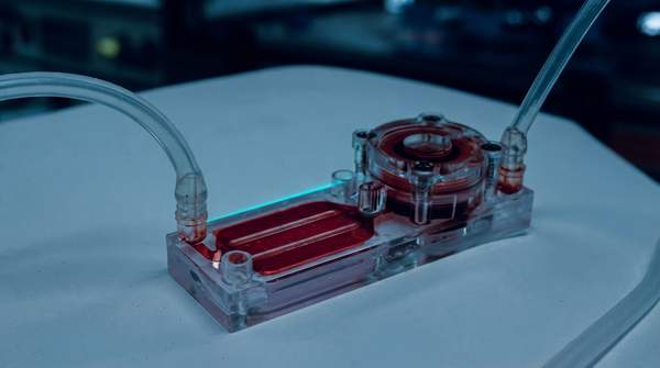
Organ-on-a-chip technology recreates the complex functions of human organs on a micro-scale, offering a more accurate and ethically sound platform for developing new drugs and personalized treatments. By combining living human cells with microfluidic engineering, these devices mimic the key cellular structures, chemical signals, and physical forces of a living organ. This provides a dynamic window into human biology that traditional laboratory models, which contribute to high drug failure rates, often fail to capture.
For decades, the path from a promising compound to an approved medicine has relied on flat, two-dimensional cell cultures and animal testing. These methods frequently fail to predict human responses, leading to costly and disappointing late-stage clinical trial failures. Organ-on-a-chip systems aim to bridge this gap by providing a more physiologically relevant testing ground. According to a 2022 article in Nature Reviews Genetics, the technology is not intended to build full-scale replacement organs but rather to replicate the smallest functional unit of an organ, allowing researchers to model diseases and test drug efficacy and toxicity with greater precision.
The Inner Workings of an Organ-on-a-Chip
At its core, an organ-on-a-chip is a microfluidic device, often made of a flexible polymer, that houses living human cells in a meticulously engineered microenvironment. The design aims to mimic the key architectural and functional features of a specific organ, such as a lung, kidney, or intestine. The use of microfluidics allows for the precise control of parameters like concentration gradients and fluid flow while consuming very small amounts of materials.
A common design, described by the company Emulate, involves two parallel, hollow channels separated by a thin, porous membrane. One channel often represents the vascular side, lined with blood vessel cells, while the other contains organ-specific cells, creating a critical tissue-vascular interface. This structure allows for crucial cell-to-cell communication and the transport of nutrients, drugs, and immune cells, much like in the human body. The cells used can be primary cells taken directly from a patient or, increasingly, induced pluripotent stem cells (iPSCs). As one research paper notes, iPSCs have become a common source for supplying human cells for various organs, enabling the creation of highly specific and even patient-derived models.
A defining feature of this technology is its ability to recreate biomechanical forces that are critical to organ function but absent in static cell cultures. For example, researchers can flow fluids through the channels to simulate blood flow, introducing a physical force known as fluid shear stress. In a lung-on-a-chip, a device first detailed in a 2010 Science article, mechanical stretching can be applied to mimic the act of breathing. This integration of a 3D extracellular matrix, biochemical factors, and biophysical cues is what allows the technology to serve as a powerful tool for disease modeling and drug screening.